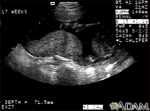
Ultrasound, normal placenta - Braxton Hicks

This is a normal ultrasound performed at 17 weeks gestation. It shows the placenta during a normal (Braxton Hicks) contraction. Throughout the pregnancy, the uterus periodically contracts to facilitate better blood flow through the placenta and the fetus. In this ultrasound, the placenta can be seen as the mound-shaped object in the middle of the screen. At the bottom of the image, the mother's vertebra can be seen as a round object. When the uterus is not contracting, the placenta would appear much flatter.

Review Date:
10/15/2024
Reviewed By: John D. Jacobson, MD, Professor Emeritus, Department of Obstetrics and Gynecology, Loma Linda University School of Medicine, Loma Linda, CA. Also reviewed by David C. Dugdale, MD, Medical Director, Brenda Conaway, Editorial Director, and the A.D.A.M. Editorial team.
Reviewed By: John D. Jacobson, MD, Professor Emeritus, Department of Obstetrics and Gynecology, Loma Linda University School of Medicine, Loma Linda, CA. Also reviewed by David C. Dugdale, MD, Medical Director, Brenda Conaway, Editorial Director, and the A.D.A.M. Editorial team.
The information provided herein should not be used during any medical emergency or for the diagnosis or treatment of any medical condition. A licensed medical professional should be consulted for diagnosis and treatment of any and all medical conditions. Links to other sites are provided for information only -- they do not constitute endorsements of those other sites. No warranty of any kind, either expressed or implied, is made as to the accuracy, reliability, timeliness, or correctness of any translations made by a third-party service of the information provided herein into any other language. © 1997-
A.D.A.M., a business unit of Ebix, Inc. Any duplication or distribution of the information contained herein is strictly prohibited.
