| Anatomy and function of the respiratory system |
The human respiratory system is divided into the upper and lower respiratory tracts.
Upper respiratory tract
The major passages and structures of the upper respiratory tract include the nose or nostrils, nasal cavity, mouth, throat (pharynx), and voice box (larynx).
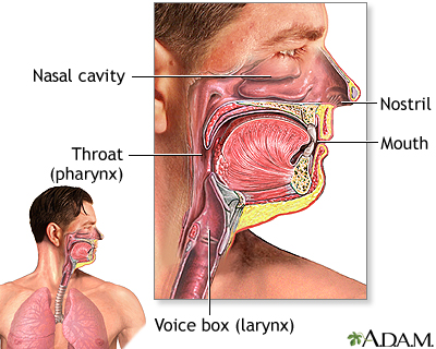
When you breathe in through your nose or mouth, the air is "filtered" through natural lines of defense that protect against illness and irritation of the respiratory tract. Nasal hairs (vibrissae) at the opening of the nostrils trap large particles of dust that might otherwise be inhaled. The entire respiratory system, as with the reproductive, digestive, and urinary systems, is lined with a mucous membrane that secretes mucus. The mucus traps smaller particles like pollen or smoke. Hairlike structures called cilia line the mucous membrane and move the particles trapped in the mucus out of the nose.
Inhaled air is moistened, warmed, and cleansed by the nasal epithelium (the tissue that lines the nasal cavity), which covers the turbinate bones (conchae) in the nasal cavity. The nasal epithelium has increased blood flow that helps to warm the inhaled air, but also facilitates nosebleeds in some people.
The pharynx is a muscular, funnel-shaped tube about 5 inches long that connects the nasal and oral cavities to the larynx. The pharynx houses the tonsils and the adenoids, which are lymphatic tissues that guard against infection by releasing white blood cells (T and B lymphocytes).
The larynx forms the entrance to the lower respiratory system. With the help of the epiglottis (a leaf-shaped flap), the larynx prevents food or liquid from entering the lower respiratory tract while swallowing. Two pairs of strong connective tissue bands that are stretched across the larynx (vocal cords) vibrate to produce sounds while talking or singing.
Lower respiratory tract
The major passages and structures of the lower respiratory tract include the windpipe (trachea) and within the lungs, the bronchi, bronchioles, and alveoli.
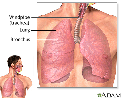
After the inhaled air moves through the larynx, it reaches the trachea. The trachea is a rigid tube about 4.5 inches long and 1 inch wide. Embedded in the walls of the trachea, C-shaped cartilage rings give the trachea rigidity and allow it to stay open all the time.
Inward airflow from the trachea then branches off to the two bronchi. One bronchus leads to the right lung, the other to the left lung. The bronchi also contain C-shaped cartilage rings like the trachea.
Deeper in the lungs, each bronchus divides into secondary and tertiary bronchi, which continue to branch to smaller airways called the bronchioles. There is no cartilage in the bronchioles, and therefore they are subject to constriction and obstruction, as during an asthma attack. The bronchioles end in air sacs called the alveoli. Alveoli are bunched together into clusters to form alveolar sacs. On the surface of each alveolus, there is a network of capillaries carrying blood that has come through veins from other parts of the body. Here gas exchange occurs -- carbon dioxide from the blood is exchanged for oxygen from the alveoli. After the blood is oxygenated, it goes to the heart (between the two lungs), where it is pumped out to all of the body tissues and extremities. When you breathe out, the carbon dioxide is exhaled and expelled from the body.
The role of the diaphragm
The diaphragm, located below the lungs, is the major muscle of respiration. It is a large, dome-shaped muscle that contracts rhythmically and continually, and most of the time, involuntarily. Upon inhalation, the diaphragm contracts and flattens and the chest cavity enlarges. This contraction creates a vacuum, which pulls air into the lungs. Upon exhalation, the diaphragm relaxes and returns to its domelike shape, and air is forced out of the lungs.
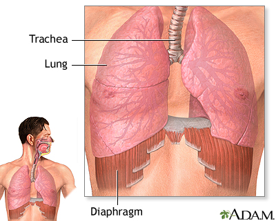
Other important respiratory muscles include the intercostal (between the ribs) and abdominal muscles.
Reviewed By: Allen J. Blaivas, DO, Clinical Assistant Professor of Medicine UMDNJ-NJMS, Attending Physician in the Division of Pulmonary, Critical Care, and Sleep Medicine, Department of Veteran Affairs, VA New Jersey Health Care System, East Orange, NJ. Review provided by VeriMed Healthcare Network. Previoulsy reviewed by David A. Kaufman, MD, Section Chief, Pulmonary, Critical Care & Sleep Medicine, Bridgeport Hospital-Yale New Haven Health System, and Assistant Clinical Professor, Yale University School of Medicine, New Haven, CT. Review provided by VeriMed Healthcare Network. (6/1/2010)
 All rights reserved.